What are leukoplakia and erythroplakia?
Leukoplakia and erythroplakia are lesions observed most frequently on the mucosa of the mouth, but also on occasion in the throat and on the vocal folds. They are commonly seen in smokers, individuals exposed to toxic irritants, and patients with nutritional deficiencies - but they also occur in the absence of such factors.
Leukoplakia and erythroplakia are generally considered precancerous lesions, although their potential to turn into cancer cannot be assessed by their appearance. This risk is assessed by examining samples under a microscope for a characteristic of cells called dysplasia. Having dysplasia does not guarantee that cells are cancerous, but it places patients at a higher risk level than the general population for eventual cancer development. Even so, the majority of leukoplakia and erythroplakia lesions, including those with dysplasia, will never lead to cancer formation.
What are the symptoms of leukoplakia and erythroplakia?
Leukoplakia and erythroplakia can go unnoticed when they occur in the mouth or the throat, and are sometimes found during a routine head and neck examination, or voice screening. When affecting the vocal folds, they will generally cause changes that can range from simple hoarseness to complete loss of voice. Symptoms typically evolve slowly.
What do leukoplakia and erythroplakia look like?
Leukoplakia means “white patch”, and erythroplakia means “red patch”, which are self-explanatory descriptions of the appearance of those lesions. Frequently, the patches appear somewhat thickened as a result of the excess keratin formation (keratosis), similar to what is seen in corns and calluses. A combination of white and red areas can be seen in the same lesion - this is known as erythroleukoplakia.
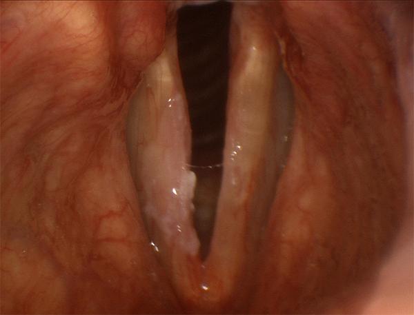
Leukoplakia has affected both vocal folds above, with predominant presence on the left fold (right side of the image). This patient is a heavy smoker who experienced voice deterioration over several years.
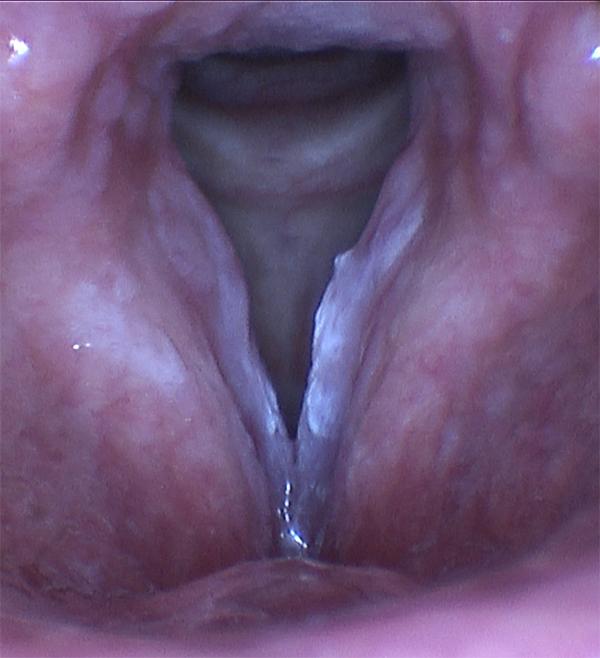
Erythroleukoplakia of the right vocal fold - this lesion combines white and red elements in a “patchwork” formation.
How are leukoplakia and erythroplakia treated?
Once leukoplakia and/or erythroplakia are identified, a biopsy and specialized examination of the specimen under a microscope are usually needed in order to determine whether the lesions harbor dysplasia. Subsequent management will depend on the location of the lesion and the nature of the biopsy findings. The options can include a combination of simple observation, laser treatments in the office, or surgical removal (microlaryngoscopy). Smoking cessation and avoidance of other irritants should always be part of the treatment strategy.
Treating leukoplakia surgically may sometimes be necessary to improve voice function when it affects the vocal folds, even in the absence of dysplasia or other concerning features on biopsy.